neurology
1) What is the evolution of the symptomatology in this patient in terms of an event timeline and where is the anatomical localization for the problem and what is the primary etiology of the patient's problem?
10 years back-Paralysis of both upper and lower limbs bilateral
1 year back-Right and left paresis due to hypokalemia
8 months backSwelling over legs
7 months back - blood infection
2 months back- neck pain
6 days back- pain along left upper limb
5 days back- chest pain, Difficulty in breathing and was able to feel her own heart beat
Anatomical localization: Cervical spine
degenerative changes that occur in the cervical spine with age.
Dehydrated disks. Disks act like cushions between the vertebrae of your spine. By the age of 40, most people's spinal disks begin drying out and shrinking, which allows more bone-on-bone contact between the vertebrae.
Bone spurs. Disk degeneration often results in the spine producing extra amounts of bone in a misguided effort to strengthen the spine. These bone spurs can sometimes pinch the spinal cord and nerve roots.
Herniated disks. Age also affects the exterior of your spinal disks. Cracks often appear, leading to bulging (herniated) disks — which sometimes can press on the spinal cord and nerve roots.
Stiff ligaments. Ligaments are cords of tissue that connect bone to bone. Spinal ligaments can stiffen with age, making your neck less flexible.
2) What are the reasons for recurrence of hypokalemia in her? Important risk factors for her hypokalemia?
Reasons for recurrence
The primary hypokalemic periodic paralysis is autosomal dominant and is exacerbated by strenuous exercise, high carbohydrate diet, cold and excitement, which was not found in this case. secondary periodic hypokalemic paralysis have been reported in association with gastroenteritis, diuretic abuse, renal tubular acidosis, Bartter syndrome, villous adenoma of colon, and hyperthyroidism.
Risk factors

Female [1] [2]
Medications like diuretics
Heart failure
Hypertension
Low BMI [3]
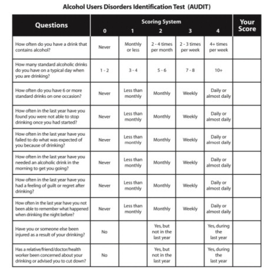
Eating disorder and alcoholism: low intake of potassium
Diarrhea, cushing syndrome, a
3) What are the changes seen in ECG in case of hypokalemia and associated symptoms?
ECG changes include flattening and inversion of T waves in mild hypokalemia, followed by Q-T interval prolongation, visible U wave and mild ST depression4 in more severe hypokalemia.